When Your Teeth Are Doing Their Best At Social Distancing
Dr. Barett Andreasen, Oral and Maxillofacial Radiologist
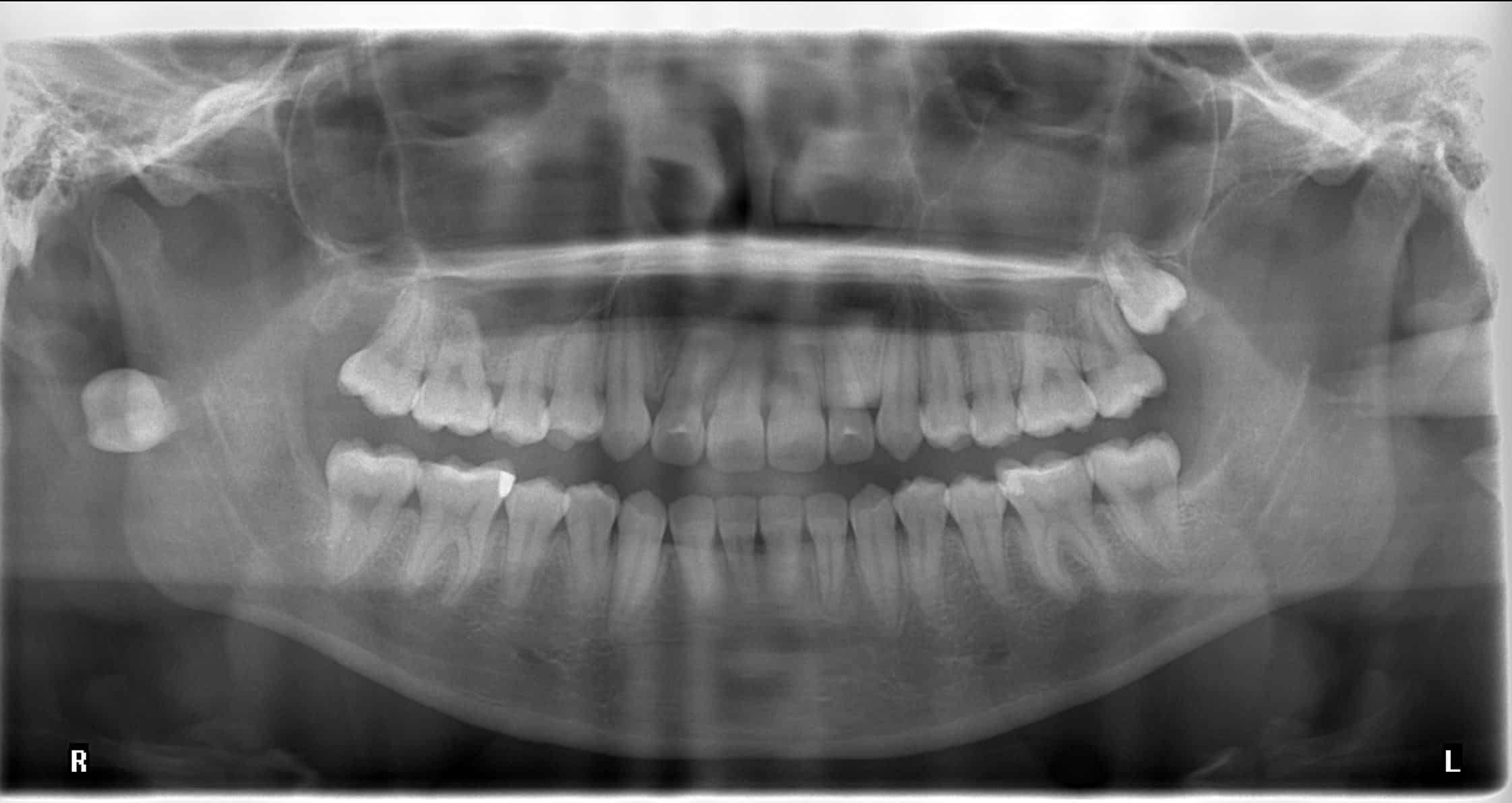
Extraction of #1 took a little detour. Where did this end up?
While we can make a fairly educated guess where the tooth is, there's a few things that can help us be more confident:
- The tooth appears larger than #16, suggesting that the tooth is magnified due to it's lingual position behind the focal trough.
- If you look closely, there's a horizontal radiopacity at the mid-level of the left posterior ramus. This is a double image of #1, again suggesting a lingual or close to mid-line position.
- Following the outline of the airway, you can make out the soft palate that is preventing the patient from swallowing/aspirating the tooth.
These three findings point to the fact that the tooth is superior to the soft palate in the nasopharyngeal airway.
Again, we could have made a fairly accurate guess on the position of the tooth. However, imagine this isn't a tooth, but a calcification or a foreign object. Using our knowledge of panoramic image formation and physics, we can more accurately diagnose our panoramic findings.
Of course, we could always just take a CBCT, but that'd just be too easy!

