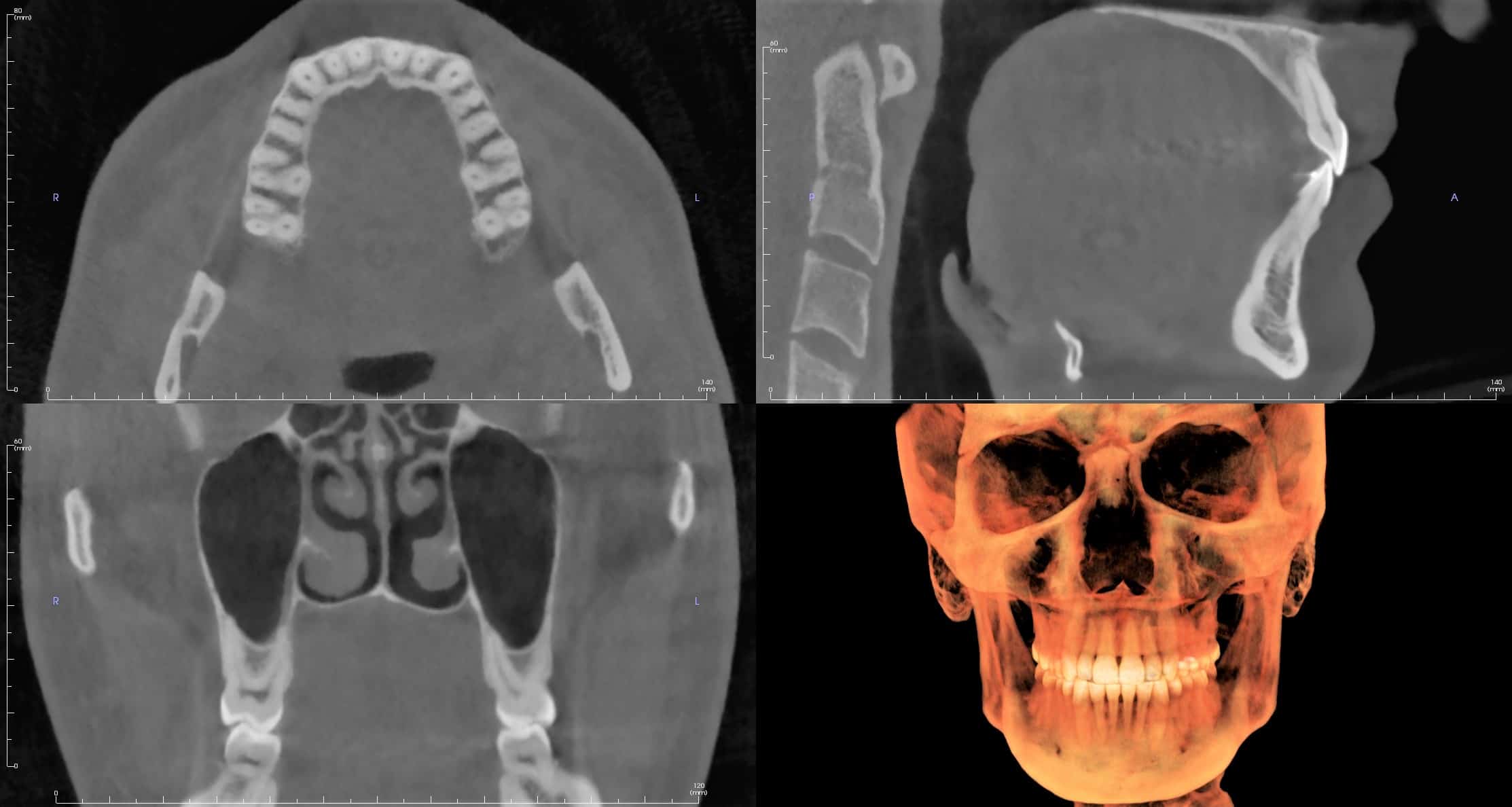
The introduction of cone-beam computed tomography (CBCT) has heralded a shift from a 2D to a 3D approach in image acquisition and patient management. This technology has given dentistry unprecedented diagnostic information that has forever changed how we approach patient treatment. But what is CBCT and how does it work?
CBCT technology is a recent technology utilizing a divergent pyramidal- or cone-shaped x-ray beam directed through the area of interest onto an x-ray detector. The x-ray source and detector rotate about the area of interest, capturing multiple projection images in a single rotation of the gantry. (This diverges from conventional CT which uses a fan-shaped x-ray beam in a helical progression to capture individual slices of the area of interest, requiring multiple passes of the gantry.)
These acquired projection images are similar to traditional 2D lateral or posterior-anterior cephs, with each subsequent image being slightly offset from the previous one. The number of these images captured normally ranges from 100 to >600 and is dependent on the number of images acquired per second, the range of the trajectory arc, and the speed of the rotation. Once the projection images have been acquired, the data must be reconstructed to create the volumetric data, which is evaluated on your favorite CBCT viewer.
Advantages of CBCT include rapid scan times, reduced patient radiation dose, high resolution images, measurements free of distortion and magnification, 3D volume rendering and the ability to reorient the scan to realign the area of interest. CBCTs are limited by artifacts (either patient-related or scanner-related), image noise, and poor soft tissue contrast.
CBCT technology has had a tremendous impact on how dentistry is practiced and will only become more commonplace as time progresses.

