Arrested Pneumatization of the Sphenoid Sinus
Dr. Barett Andreasen, Oral and Maxillofacial Radiologist
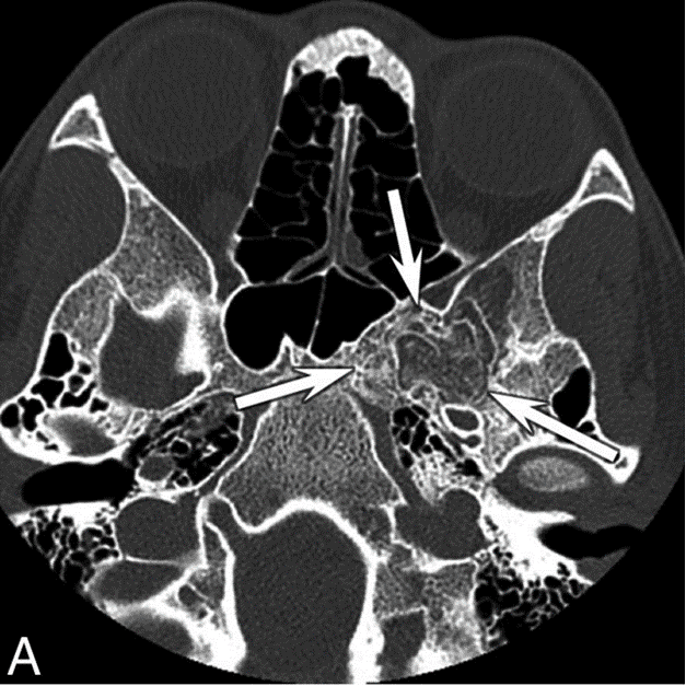
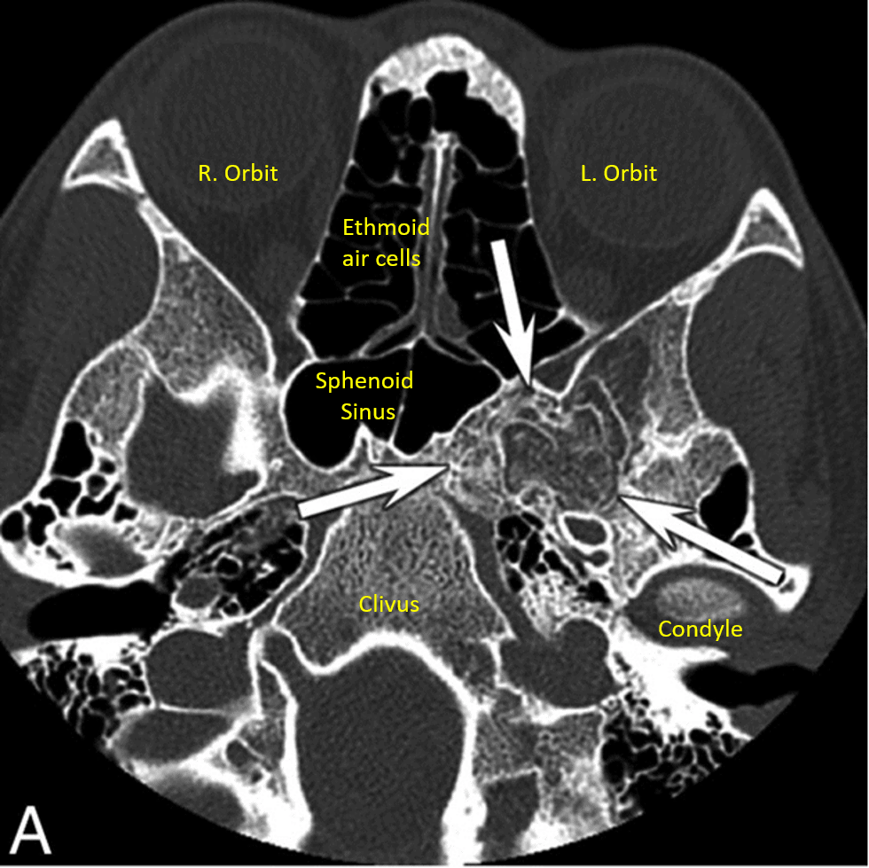
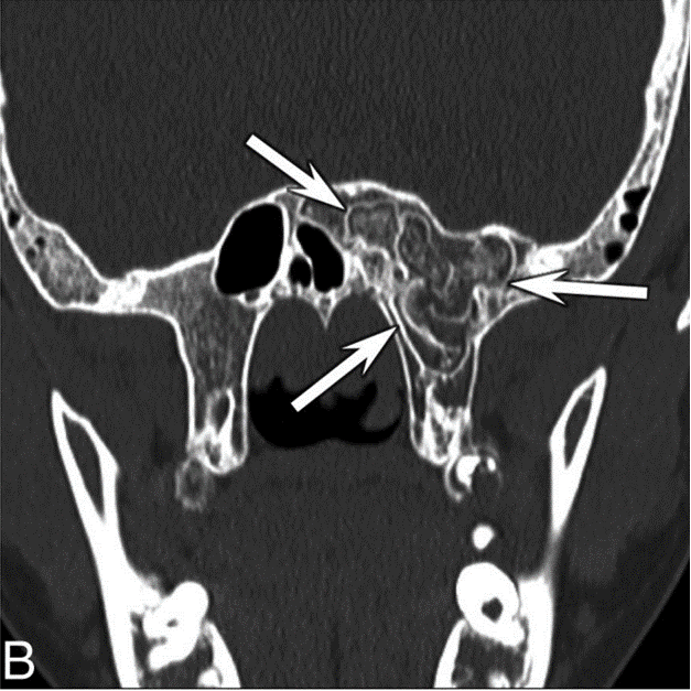
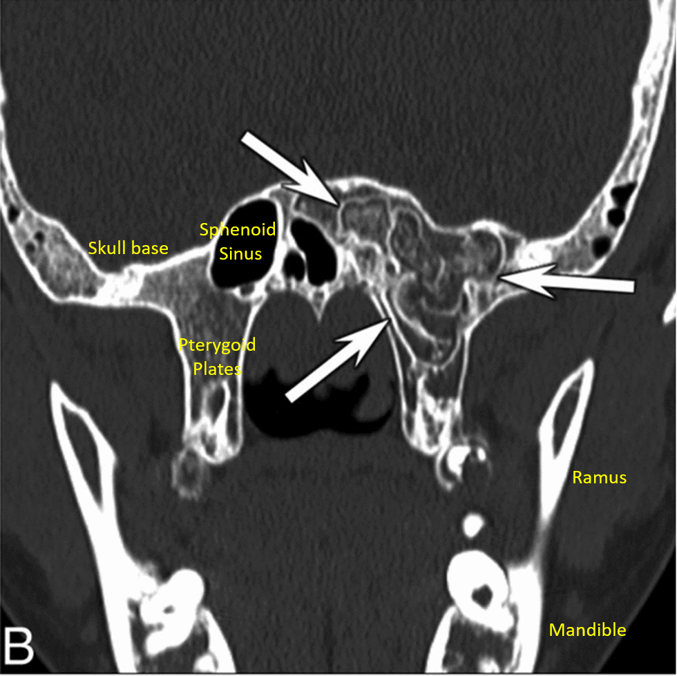
Today I'd like to share an interesting case in a region that is frequently captured in dental CBCT scans. I've included images with labels of the surrounding anatomy as well as a skull with approximate locations of the slices to help you get oriented.
A patient presented with an incidental finding located at the left sphenoid sinus and greater wing of the sphenoid. The area of interest is delineated by white arrows in the images. The CT slices show a nonexpansile lesion with a thin cortical margin and curvilinear internal calcifications. Patient denied any symptoms.
While the appearance is very unusual, this finding represents arrested pneumatization of the sphenoid sinus, a normal anatomical variation caused by an interruption of the normal development of the sinuses. The development of the sphenoid sinus begins with fatty transformation and fat involution of the bone marrow, followed by aeration of the marrow that results in pneumatization of the bone. The normal pneumatization process occurs soon after birth and ends at 10-14 years of age. Interruption of this process results in an area of persistent atypical fatty marrow that persists into adulthood and manifests as a nonexpansile lesion with thin cortical margins and curvilinear internal calcifications. The lesion will also respect the margins of adjacent foramina and lacks a normal trabecular pattern found in the adjacent bone. While arrested pneumatization can technically occur in any sinus, there is a marked predominance for the sphenoid sinus; however, the reasons why such a predominance is present or the causes for the alteration in normal development are not fully understood.
Potential differential diagnoses include: intraosseous lipoma, intraosseous hemangioma, fibrous dysplasia, ossifying fibroma, chordoma, chondrosarcoma, and metastases. In contrast to arrested pneumatization, all these conditions usually show signs of mass effect on the surrounding structures.
As this is a normal anatomical variation, no treatment is necessary. However, MR imaging may be indicated to rule out osseous pathology.
This case came from the article "The CT Prevalence of Arrested Pneumatization of the sphenoid Sinus in Patients with Sickle Cell Disease" by A.V. Prabhu, and B.F. Branstetter.


